Reactivity
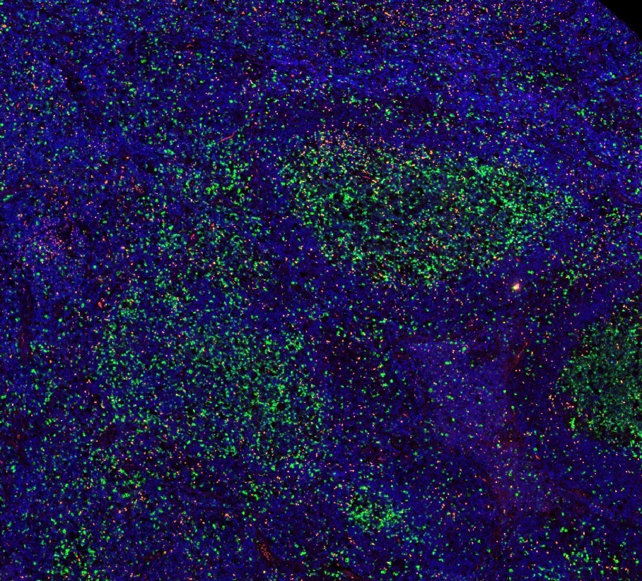
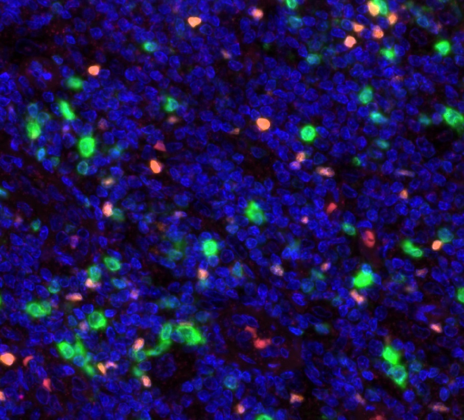
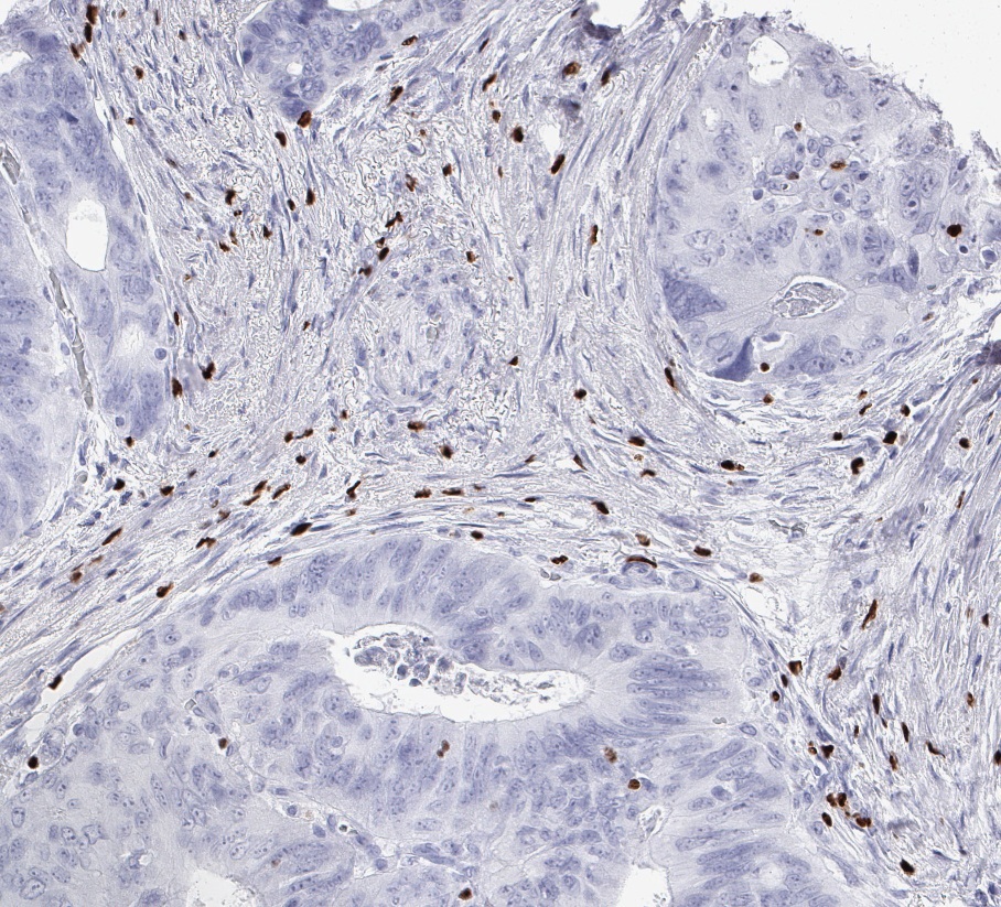
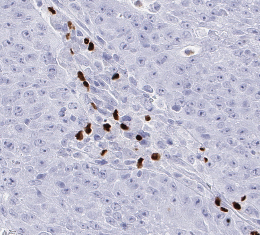
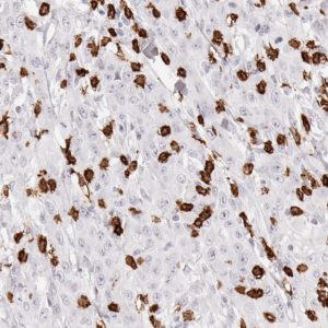
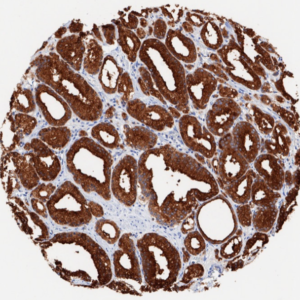
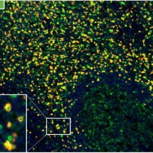
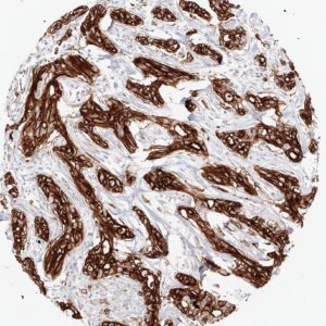
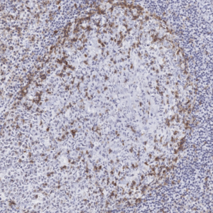
Clone FX3 has been developed and validated for fluorescence multiplex IHC studies of FOXP3 expression in human tissues.
FOXP3 (Forkhead box protein P3) is mainly expressed in Regulatory T (Treg) cells, a subset of CD4+ T-cells, that play a suppressive role in the immune system. Treg cells ensure immune homeostasis through their ability to suppress the activation and function of leukocytes. FOXP3 has emerged as a prominent target for the development of new immunotherapies for cancer and autoimmune diseases.
IHC protocols
Staining protocols for anti-human FOXP3 antibody clone FX3
| Cat.No.: |
DIA-FX3 |
| Isotype: |
Mouse IgG2a/k |
| Specificity: |
Human FOXP3 |
| Physical State: |
Lyophilized powder |
| Reconstitution: |
DIA-FX3, restore to 100µl. Reconstitute with sterile destilled water by gentle shaking for 10 minutes. |
Manual stain with autoclave
- Pretreatment buffer: 121°C / 5 min / pH7,8
- Incubation primary antibody: 60 min / 37°C (Dilution: 1:100)
- Envision HRP rabbit/mouse: 30 min / 37°C
Applicaple for automated staining procedures and validated for mulitcolor immunofluorescebnce (multiplexed IHC).
References
Scientific publications for anti-FOXP3 clone FX3 have been submitted and reference data will be available soon.

