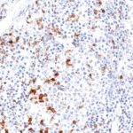
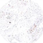
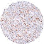
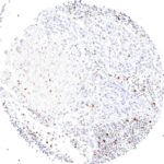
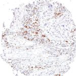
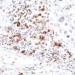
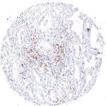
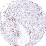
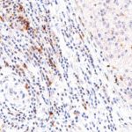
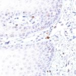
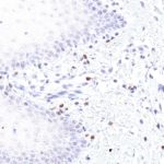
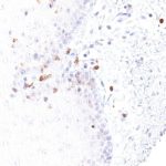
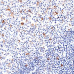
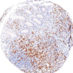
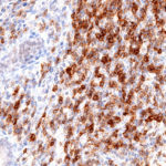
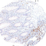
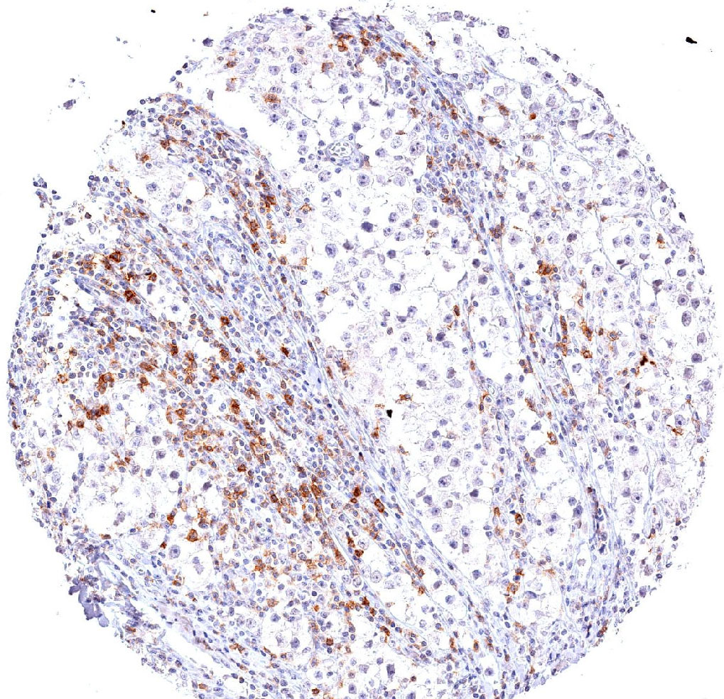
Antibody clones TG1 and TG2 for studying invading TIGIT-positive T cells in FFPE tissues
TIGIT antibody
TIGIT antibody
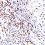
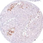
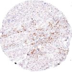
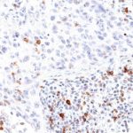
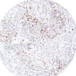
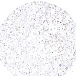
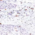
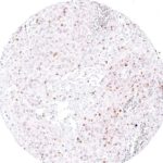
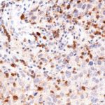
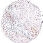
Strong TIGIT expression in a large number of tumor infiltrating lymphocytes in a seminoma with dense lymphocytic infiltrate
TIGIT antibody
TIGIT antibody
TIGIT antibody
TIGIT antibody
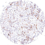
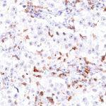
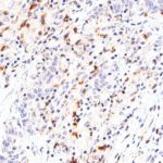
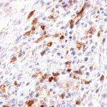
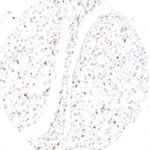
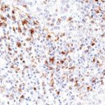
Strong TIGIT expression in tumor infiltrating lymphocytes in a seminoma with sparse lymphocytic infiltrate.
Fig. 4: Strong TIGIT expression in tumor infiltrating lymphocytes in a seminoma with sparse lymphocytic infiltrate.
TIGIT antibody
TIGIT antibody
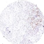
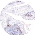
Fig. 6: High fraction of TIGIT positive cells in peritumoral lymphocytes in a case of penis carcinoma.
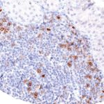
Fig. 7: Low grade neuroendocrine carcinoma with scattered infiltrate containing TIGIT positive lymphocytes.
TIGIT antibody
TIGIT antibody
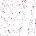
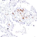
Fig. 9: High grade neuroendocrine carcinoma with focal accumulation of TIGIT positive lymphocytes.
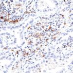
Fig. 10: High grade neuroendocrine carcinoma with focal accumulation of TIGIT positive lymphocytes.
Fig. 11: Dense peritumoral and intratumoral infiltrate of TIGIT positive lymphocytes in a case of a poorly differentiated squamous cell carcinoma of the esophagus.
Fig. 12: High frequency of TIGIT positive lymphocytes in a lymphoepithelial carcinoma.
TIGIT antibody
TIGIT antibody
TIGIT antibody
TIGIT antibody
TIGIT antibody
TIGIT antibody
TIGIT antibody
TIGIT antibody
Invasive urothelial carcinoma with dense peritumoral and intratumoral infiltrate of TIGIT positive lymphocytes.
TIGIT antibody
TIGIT antibody
TIGIT antibody
TIGIT antibody
TIGIT antibody
TIGIT antibody
High frequency of TIGIT positive lymphocytes in a Hodgkin lymphoma.
Fig. 20: High frequency of TIGIT positive lymphocytes in a Hodgkin lymphoma.
Fig. 21: TIGIT positive lymphocytes are present in the peritumoral and intratumoral lymphocytic infiltrate of a squamous cell carcinoma of the lung.
Fig. 22: TIGIT positive lymphocytes are present in the peritumoral and intratumoral lymphocytic infiltrate of a squamous cell carcinoma of the lung.
TIGIT antibody
TIGIT antibody
Fig. 24: High frequency of TIGIT positive intratumoral lymphocytes in a squamous cell carcinoma of the lung.
TIGIT antibody
TIGIT antibody
Fig. 26: TIGIT positive lymphocytes are located peritumoral but not intratumoral in a squamous cell ca
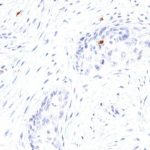
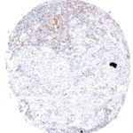
Fig. 27: Sparse TIGIT positive lymphocytes in a squamous cell carcinoma of the lung with strong desmoplastic reaction.
Fig. 28: Sparse TIGIT positive lymphocytes in a squamous cell carcinoma of the lung with strong desmoplastic reaction.
Fig. 29: High density of TIGIT positive lymphocytes in a squamous cell carcinoma of the lung.
TIGIT antibody
TIGIT antibody
High density of TIGIT positive lymphocytes in a squamous cell carcinoma of the lung.
TIGIT antibody
TIGIT antibody
Fig. 32: TIGIT positive lymphocytes in a bronchioloalveolar carcinoma of the lung.
Fig. 33: High frequency of TIGIT positive lymphocytes in an adenocarcinoma of the lung.
TIGIT antibody
TIGIT antibody
TIGIT antibody
TIGIT antibody
Fig. 36: High frequency of peritumoral TIGIT positive lymphocytes in a large cell adenocarcinoma of the lung.
Fig. 37: Abundant TIGIT positive lymphocytes in an adenocarcinoma of the ampulla vateri.
TIGIT antibody
TIGIT antibody
TIGIT antibody
TIGIT antibody
High number of TIGIT positive lymphocytes in an angiosarcoma
TIGIT antibody
TIGIT antibody
High number of TIGIT positive lymphocytes in an angiosarcoma.
Fig. 41: Frequent TIGIT positive lymphocytes in a liposarcoma.
Fig. 42: Frequent TIGIT positive lymphocytes in a liposarcoma.
Fig. 43: TIGIT positive lymphocytes are abundant in a Whartin tumor.
TIGIT antibody
TIGIT antibody
TIGIT positive lymphocytes are abundant in a Whartin tumor.
Fig. 45: Dense infiltration of an ovarian carcinoma by TIGIT positive lymphocytes.
Fig. 46: Dense infiltration of an ovarian carcinoma by TIGIT positive lymphocytes.
Fig. 47: A cervix uteri squamous cell carcinoma with numerous intratumoral TIGIT positive lymphocytes.
Fig. 48: A cervix uteri squamous cell carcinoma with numerous intratumoral TIGIT positive lymphocytes.
Fig 49: Squamous cell carcinoma of the uterine cervix with peritumoral and intratumoral infiltrate of TIGIT positive lymphocytes.
Fig. 50: Fig 49: Squamous cell carcinoma of the uterine cervix with peritumoral and intratumoral infiltrate of TIGIT positive lymphocytes.
Fig. 51: Squamous cell carcinoma of the uterine cervix with abundant intratumoral infiltrate of TIGIT positive lymphocytes.
Fig. 52: Squamous cell carcinoma of the uterine cervix with abundant intratumoral infiltrate of TIGIT positive lymphocytes.
Fig. 53: Squamous cell carcinoma of the uterine cervix with a high number of intratumoral TIGIT positive lymphocytes.
Fig. 54: Squamous cell carcinoma of the uterine cervix with a high number of intratumoral TIGIT positive lymphocytes.
Fig. 55: Peritumoral TIGIT positive lymphocytes in a case of endometrium carcinoma.
Fig. 56: Peritumoral TIGIT positive lymphocytes in a case of endometrium carcinoma.
Fig. 57: High density of TIGIT positive lymphocytes in a medullary breast cancer.
Fig. 58: High density of TIGIT positive lymphocytes in a medullary breast cancer.
High density of TIGIT positive lymphocytes in a medullary breast cancer.
Fig. 59: A case of medullary breast cancer with abundant TIGIT positive lymphocytes.
Fig. 60: A case of medullary breast cancer with abundant TIGIT positive lymphocytes.
Fig. 61: High density of peritumoral and intratumoral TIGIT positive lymphocytes in a medullary breast cancer.
Fig. 62: High density of peritumoral and intratumoral TIGIT positive lymphocytes in a medullary breast cancer.
Fig. 63: TIGIT positive lymphocytes in the subepithelial tissue of the lip (oral epithelium)
Fig. 64: TIGIT positive lymphocytes in the subepithelial tissue of the lip (oral epithelium)
Fig. 65: Oral mucosa with TIGIT positive lymphocytes in the subepithelial stroma.
Fig. 66: Oral mucosa with TIGIT positive lymphocytes in the subepithelial stroma.
TIGIT antibody
TIGIT antibody
Fig. 68: Oral mucosa with intraepithelial and subepithelial TIGIT positive lymphocytes.
Fig. 69: Abundant TIGIT positive lymphocytes in the spleen.
Fig. 70: Abundant TIGIT positive lymphocytes in the spleen.
Fig. 71: TIGIT positive lymphocytes are frequent in the thymus.
Fig. 72: TIGIT positive lymphocytes are frequent in the thymus.
Fig. 73: Thymus with high number of TIGIT positive lymphocytes.
Fig. 74: Thymus with high number of TIGIT positive lymphocytes.
Fig. 75: TIGIT positive lymphocytes are abundant in the mucosa of the appendix.
Fig. 76: TIGIT positive lymphocytes are abundant in the mucosa of the appendix.
Fig. 78: Appendix mucosa with dense accumulation of TIGIT positive lymphocytes.
Fig. 79: Appendix mucosa with dense accumulation of TIGIT positive lymphocytes.
Fig. 80: Appendix mucosa with TIGIT positive lymphocytes.
Fig. 81: Appendix mucosa with TIGIT positive lymphocytes.
TIGIT antibody

TIGIT at the Immune Checkpoint

A growing number of immune checkpoints develop as targets for anticancer therapy. Cancer immunology studies have shown that cancer cells together with cells of the surrounding microenvironment generate co-inhibitory signals by upregulating the expression of components which suppress the antitumor immune response. The TIGIT pathway interacts with different inhibitory checkpoint pathways. TIGIT provides significant promise for immunotherapy, especially in combination with other immune checkpoint inhibitors.

The immunoreceptor TIGIT (T-cell immunoreceptor with Ig and ITIM domains) is a member of the poliovirus receptor (PVR) family.  Expression of TIGIT has been reported on NK cells, regulatory T cells, follicular T helper cells, memory CD4+ T cells, and CD8+ T cells, but TIGIT is not expressed on B cells or naive CD4+ T cells. TIGIT acts as an inhibitory immune checkpoint on both T cells and natural killer (NK) cells by a highly complex pathway. Known ligands for TIGIT include CD155 and CD112. Moreover, the TIGIT/CD155/CD112 network also interacts with further checkpoint regulators.

In inflammation and in multiple cancer models T cells have been shown to upregulate TIGIT expression. In several types of cancer, the ligands CD155 and CD112 are also highly expressed on dendritic cells and macrophages. Moreover, TIGIT expression is highly correlated with the expression of other co-inhibitory molecules, including PD-1. In addition to directly inhibiting cytotoxic T-cell activity, TIGIT can stimulate an immunosuppressive microenvironment by influencing other immune cells: For example, TIGIT binds CD155 on the surface of dendritic cells or manipulates NK cell activity. Drugs inhibiting TIGIT activity are currently being developed.

Antibody clone TG1

Clone TG1 is the first monoclonal antibody for the immunohistochemical (IHC) detection of TIGIT (T cell immunoreceptor with Ig and ITIM domains) in routine FFPE human tissue specimen. Furthermore TG1 has been validated for the identification of TIGIT positive T-cells infiltrating human tumors with the aim to allow a detection of TIGIT in the tumor microenvironment by IHC. Immunohistochemical (IHC) application of monoclonal antibody TG1 may provide valuable information for clinical research and potential therapeutic interventions specifically targeting the TIGIT-related tumor immunology checkpoint.

New – clone TG2

The newly developed clone TG2 has a higher affinity to the target protein TIGIT and shows a stronger staining intensity in IHC studies on tonsil when compared to TG1.

  • Higher affinity as compared to clone TG1 (tested on tonsil)
  • Stronger staining intensity
  • Higher Dilutability

1.
Li W et al. Expression of the immune checkpoint receptor TIGIT in Hodgkin’s lymphoma. BMC Cancer 2018, 18: 1209.
doi.org/10.1186/s12885-018-5111-1
2.
Blessin NC et al. Patterns of TIGIT expression in normal lymphatic tissue, inflammation and cancer. Disease Markers 2019, Jan 10;2019:5160565. eCollection 2019.
doi.org/10.1155/2019/5160565
3.
Hinsch A et al. et al. Expression of the immune checkpoint receptor TIGIT in seminoma. Oncol Lett. 2019, 18: 1497–1502.
doi: 10.3892/ol.2019.10428
4.
Scimeca, M. et al. Programmed death ligand 1 expression in prostate cancer cells is associated with deep changes of the tumor inflammatory infiltrate composition. Urol. Oncol. 2019, 37, 297.e19-297.e31.
doi: 10.1016/j.urolonc.2019.02.013
5.
Annibali, O., Bianchi, A., Grifoni, A. et al. A novel scoring system for TIGIT expression in classic Hodgkin lymphoma. Sci Rep 11, 7059, 2021.
doi: 10.1038/s41598-021-86655-8
6.
Murakami D et al. Prognostic value of CD155/TIGIT expression in patients with colorectal cancer. PLoS ONE 17(3), 2022.
doi.org/10.1371/journal.pone.0265908
7.
Niebel D et al. DNA methylation regulates TIGIT expression within the melanoma microenvironment, is prognostic for overall survival, and predicts progression‑free survival in patients treated with anti‑PD‑1 immunotherapy. Clinical Epigenetics 14:50, 2022.
doi.org/10.1186/s13148-022-01270-2

TIGIT Antibody Productdetails

Menu