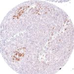
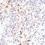
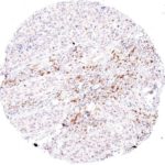
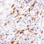
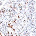
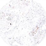
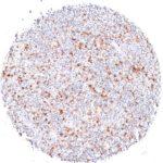
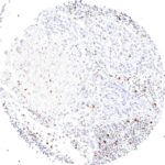
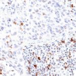
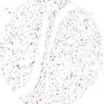
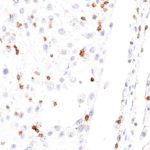
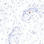
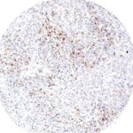
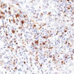
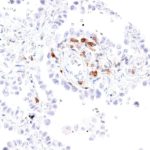
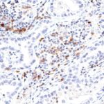
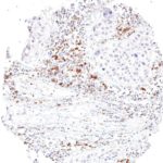
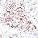
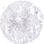
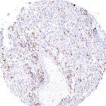
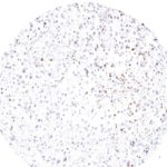
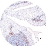
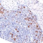
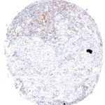
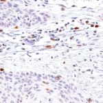
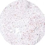
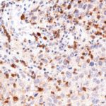
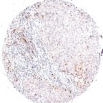
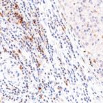
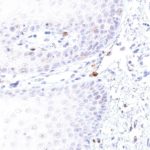
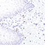
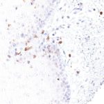
Antikörperklone TG1 und TG2 für den Nachweis TIGIT-positiver T-Zellen in FFPE Gewebe
TIGIT antibody
TIGIT antibody
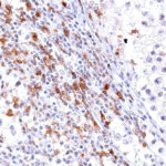
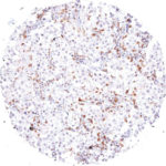
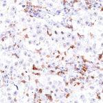
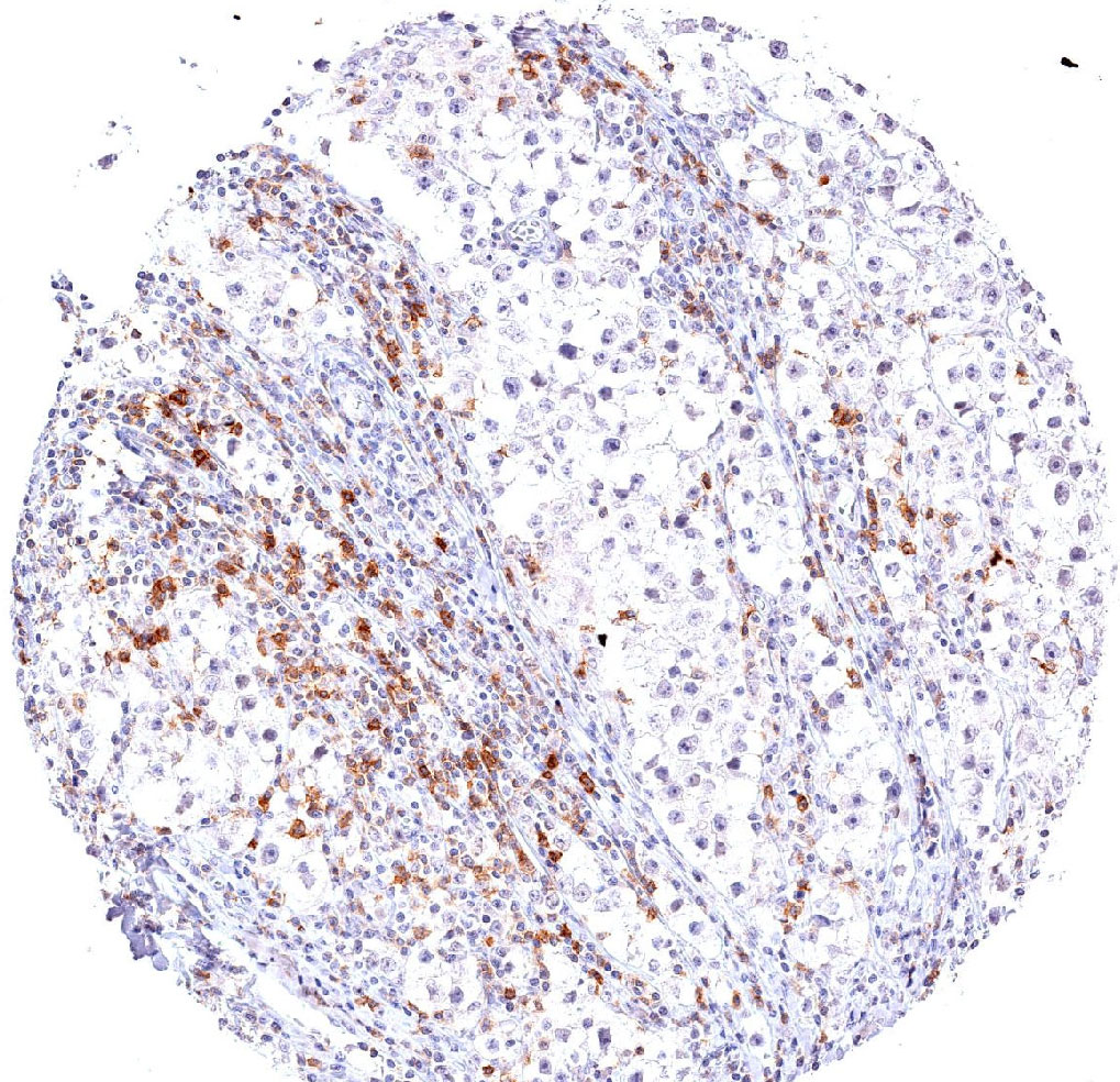
Strong TIGIT expression in a large number of tumor infiltrating lymphocytes in a seminoma with dense lymphocytic infiltrate
TIGIT antibody
TIGIT antibody
TIGIT antibody
TIGIT antibody
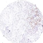
Strong TIGIT expression in tumor infiltrating lymphocytes in a seminoma with sparse lymphocytic infiltrate.
Fig. 4: Strong TIGIT expression in tumor infiltrating lymphocytes in a seminoma with sparse lymphocytic infiltrate.
TIGIT antibody
TIGIT antibody
Fig. 6: High fraction of TIGIT positive cells in peritumoral lymphocytes in a case of penis carcinoma.
Fig. 7: Low grade neuroendocrine carcinoma with scattered infiltrate containing TIGIT positive lymphocytes.
TIGIT antibody
TIGIT antibody
Fig. 9: High grade neuroendocrine carcinoma with focal accumulation of TIGIT positive lymphocytes.
Fig. 10: High grade neuroendocrine carcinoma with focal accumulation of TIGIT positive lymphocytes.
Fig. 11: Dense peritumoral and intratumoral infiltrate of TIGIT positive lymphocytes in a case of a poorly differentiated squamous cell carcinoma of the esophagus.
Fig. 12: High frequency of TIGIT positive lymphocytes in a lymphoepithelial carcinoma.
TIGIT antibody
TIGIT antibody
TIGIT antibody
TIGIT antibody
TIGIT antibody
TIGIT antibody
TIGIT antibody
TIGIT antibody
Invasive urothelial carcinoma with dense peritumoral and intratumoral infiltrate of TIGIT positive lymphocytes.
TIGIT antibody
TIGIT antibody
TIGIT antibody
TIGIT antibody
TIGIT antibody
TIGIT antibody
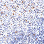
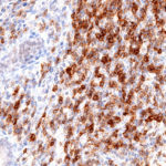
High frequency of TIGIT positive lymphocytes in a Hodgkin lymphoma.
Fig. 20: High frequency of TIGIT positive lymphocytes in a Hodgkin lymphoma.
Fig. 21: TIGIT positive lymphocytes are present in the peritumoral and intratumoral lymphocytic infiltrate of a squamous cell carcinoma of the lung.
Fig. 22: TIGIT positive lymphocytes are present in the peritumoral and intratumoral lymphocytic infiltrate of a squamous cell carcinoma of the lung.
TIGIT antibody
TIGIT antibody
Fig. 24: High frequency of TIGIT positive intratumoral lymphocytes in a squamous cell carcinoma of the lung.
TIGIT antibody
TIGIT antibody
Fig. 26: TIGIT positive lymphocytes are located peritumoral but not intratumoral in a squamous cell ca
Fig. 27: Sparse TIGIT positive lymphocytes in a squamous cell carcinoma of the lung with strong desmoplastic reaction.
Fig. 28: Sparse TIGIT positive lymphocytes in a squamous cell carcinoma of the lung with strong desmoplastic reaction.
Fig. 29: High density of TIGIT positive lymphocytes in a squamous cell carcinoma of the lung.
TIGIT antibody
TIGIT antibody
High density of TIGIT positive lymphocytes in a squamous cell carcinoma of the lung.
TIGIT antibody
TIGIT antibody
Fig. 32: TIGIT positive lymphocytes in a bronchioloalveolar carcinoma of the lung.
Fig. 33: High frequency of TIGIT positive lymphocytes in an adenocarcinoma of the lung.
TIGIT antibody
TIGIT antibody
TIGIT antibody
TIGIT antibody
Fig. 36: High frequency of peritumoral TIGIT positive lymphocytes in a large cell adenocarcinoma of the lung.
Fig. 37: Abundant TIGIT positive lymphocytes in an adenocarcinoma of the ampulla vateri.
TIGIT antibody
TIGIT antibody
TIGIT antibody
TIGIT antibody
High number of TIGIT positive lymphocytes in an angiosarcoma
TIGIT antibody
TIGIT antibody
High number of TIGIT positive lymphocytes in an angiosarcoma.
Fig. 41: Frequent TIGIT positive lymphocytes in a liposarcoma.
Fig. 42: Frequent TIGIT positive lymphocytes in a liposarcoma.
Fig. 43: TIGIT positive lymphocytes are abundant in a Whartin tumor.
TIGIT antibody
TIGIT antibody
TIGIT positive lymphocytes are abundant in a Whartin tumor.
Fig. 45: Dense infiltration of an ovarian carcinoma by TIGIT positive lymphocytes.
Fig. 46: Dense infiltration of an ovarian carcinoma by TIGIT positive lymphocytes.
Fig. 47: A cervix uteri squamous cell carcinoma with numerous intratumoral TIGIT positive lymphocytes.
Fig. 48: A cervix uteri squamous cell carcinoma with numerous intratumoral TIGIT positive lymphocytes.
Fig 49: Squamous cell carcinoma of the uterine cervix with peritumoral and intratumoral infiltrate of TIGIT positive lymphocytes.
Fig. 50: Fig 49: Squamous cell carcinoma of the uterine cervix with peritumoral and intratumoral infiltrate of TIGIT positive lymphocytes.
Fig. 51: Squamous cell carcinoma of the uterine cervix with abundant intratumoral infiltrate of TIGIT positive lymphocytes.
Fig. 52: Squamous cell carcinoma of the uterine cervix with abundant intratumoral infiltrate of TIGIT positive lymphocytes.
Fig. 53: Squamous cell carcinoma of the uterine cervix with a high number of intratumoral TIGIT positive lymphocytes.
Fig. 54: Squamous cell carcinoma of the uterine cervix with a high number of intratumoral TIGIT positive lymphocytes.
Fig. 55: Peritumoral TIGIT positive lymphocytes in a case of endometrium carcinoma.
Fig. 56: Peritumoral TIGIT positive lymphocytes in a case of endometrium carcinoma.
Fig. 57: High density of TIGIT positive lymphocytes in a medullary breast cancer.
Fig. 58: High density of TIGIT positive lymphocytes in a medullary breast cancer.
High density of TIGIT positive lymphocytes in a medullary breast cancer.
Fig. 59: A case of medullary breast cancer with abundant TIGIT positive lymphocytes.
Fig. 60: A case of medullary breast cancer with abundant TIGIT positive lymphocytes.
Fig. 61: High density of peritumoral and intratumoral TIGIT positive lymphocytes in a medullary breast cancer.
Fig. 62: High density of peritumoral and intratumoral TIGIT positive lymphocytes in a medullary breast cancer.
Fig. 63: TIGIT positive lymphocytes in the subepithelial tissue of the lip (oral epithelium)
Fig. 64: TIGIT positive lymphocytes in the subepithelial tissue of the lip (oral epithelium)
Fig. 65: Oral mucosa with TIGIT positive lymphocytes in the subepithelial stroma.
Fig. 66: Oral mucosa with TIGIT positive lymphocytes in the subepithelial stroma.
TIGIT antibody
TIGIT antibody
Fig. 68: Oral mucosa with intraepithelial and subepithelial TIGIT positive lymphocytes.
Fig. 69: Abundant TIGIT positive lymphocytes in the spleen.
Fig. 70: Abundant TIGIT positive lymphocytes in the spleen.
Fig. 71: TIGIT positive lymphocytes are frequent in the thymus.
Fig. 72: TIGIT positive lymphocytes are frequent in the thymus.
Fig. 73: Thymus with high number of TIGIT positive lymphocytes.
Fig. 74: Thymus with high number of TIGIT positive lymphocytes.
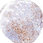
Fig. 75: TIGIT positive lymphocytes are abundant in the mucosa of the appendix.
Fig. 76: TIGIT positive lymphocytes are abundant in the mucosa of the appendix.
Fig. 78: Appendix mucosa with dense accumulation of TIGIT positive lymphocytes.
Fig. 79: Appendix mucosa with dense accumulation of TIGIT positive lymphocytes.
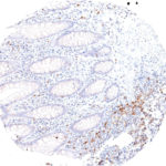
Fig. 80: Appendix mucosa with TIGIT positive lymphocytes.
Fig. 81: Appendix mucosa with TIGIT positive lymphocytes.
TIGIT antibody

TIGIT im Immuncheckpoint

Immuncheckpoint-Proteine bieten vielversprechende Ansätze für die Krebstheraphie. Krebszellen können zusammen mit Zellen aus der Mikroumgebung des Tumors die körpereigene Immunabwehr unterdrücken, indem sie lokal immunhemmende Komponeten heraufregeln. Der TIGIT-Signalweg spielt eine wichtige Rolle bei der Hemmung der Immunantwort und verknüpft verschiedene Immuncheckpoints. Die neuen immunotherapeutische Ansätze setzen daher auf die Hemmung der TIGIT-Aktivität, insbesondere in einer Kombination mit weiteren Immuncheckpoint-Hemmern.

Der Immunrezeptor TIGIT (T-Zell-Immunrezeptor mit Ig- und ITIM-Domänen) gehört zur Familie der Poliovirus-Rezeptoren (PVR). TIGIT wird auf NK-Zellen, regulatorischen T-Zellen, follikulären T-Helferzellen, Gedächtnis-CD4 + T-Zellen und CD8 + T-Zellen exprimiert, aber nicht auf B-Zellen oder naiven CD4 + T-Zellen. TIGIT wirkt dabei auf einem hochkomplexen Weg als inhibitorischer Immuncheckpoint sowohl auf T-Zellen als auch auf natürliche Killerzellen (NK). Zu den bekannten Liganden für TIGIT gehören CD155 und CD112. Darüberhinaus interagiert das TIGIT / CD155 / CD112-Netzwerk mit weiteren Checkpoint-Reglern.

Studien an Entzündungs- und Krebsmodellen zeigen, dass T-Zellen die TIGIT-Expression hochregulieren können. Auch die TIGIT-Liganden CD155 und CD112 werden auf dendritischen Zellen und Makrophagen bei verschiedenen Krebsarten stark exprimiert. Darüberhinaus korreliert die TIGIT-Expression stark mit der Expression anderer co-inhibitorischer Moleküle, einschließlich PD-1. Zusätzlich zur direkten Hemmung zytotoxischer T-Zellen kann TIGIT weitere Immunzellen beeinflussen, um eine immunsupressive Tumorumgebung zu stimulieren. TIGIT bindet beispielsweise auf der Oberfläche dendritischer Zellen CD155 und hat einen Einfluß auf die Aktivität von NK-Zellen. Es sind zahlreiche Arzneimittel in der Entwicklung, die die Aktivität von TIGIT hemmen.

Antikörperklon TG1

Der Klon TG1 ist der erste monoklonale Antikörper für den immunhistochemischen (IHC) Nachweis von TIGIT (T-Zell-Immunrezeptor mit Ig- und ITIM-Domänen) in standardmäßig Formalin-fixierten und Paraffin-eingebetteten Gewebeproben. TG1 wurde auf zahlreichen Tumoren für den IHC-Nachweis TIGIT-positiver T-Zellen in der Tumormikroumgebung validiert. Histologische Studien mit TG1 an Tumorgeweben können wertvolle Hinweise für die klinische Forschung und für therapeutische Interventionen liefern, die am TIGIT-Tumorimmunologie-Checkpoint ansetzen.

Neu – Klon TG2

Der neu entwickelte Klon TG2 hat eine höhere Affinität zum Zielprotein TIGIT und zeigt in IHC-Vergleichstudien mit Klon TG1 auf Tonsille eine stärkere Färbeintensität.

  • Höhere Affinität im Vergleich zu dem Vorgänger TG1 (auf Tonsille getestet)
  • Stärkeres Färbesignal
  • Höhere Verdünnbarkeit

1.
Li W et al. Expression of the immune checkpoint receptor TIGIT in Hodgkin’s lymphoma. BMC Cancer 2018, 18: 1209.
doi.org/10.1186/s12885-018-5111-1
2.
Blessin NC et al. Patterns of TIGIT expression in normal lymphatic tissue, inflammation and cancer. Disease Markers 2019, Jan 10;2019:5160565. eCollection 2019.
doi.org/10.1155/2019/5160565
3.
Hinsch A et al. et al. Expression of the immune checkpoint receptor TIGIT in seminoma. Oncol Lett. 2019, 18: 1497–1502.
doi: 10.3892/ol.2019.10428
4.
Scimeca, M. et al. Programmed death ligand 1 expression in prostate cancer cells is associated with deep changes of the tumor inflammatory infiltrate composition. Urol. Oncol. 2019, 37, 297.e19-297.e31.
doi: 10.1016/j.urolonc.2019.02.013
5.
Annibali, O., Bianchi, A., Grifoni, A. et al. A novel scoring system for TIGIT expression in classic Hodgkin lymphoma. Sci Rep 11, 7059, 2021.
doi: 10.1038/s41598-021-86655-8
6.
Murakami D et al. Prognostic value of CD155/TIGIT expression in patients with colorectal cancer. PLoS ONE 17(3), 2022.
doi.org/10.1371/journal.pone.0265908
7.
Niebel D et al. DNA methylation regulates TIGIT expression within the melanoma microenvironment, is prognostic for overall survival, and predicts progression‑free survival in patients treated with anti‑PD‑1 immunotherapy. Clinical Epigenetics 14:50, 2022.
doi.org/10.1186/s13148-022-01270-2

TIGIT Antikörper Produktdetails

Menü